CYTOGENOMIC TESTS.
Genomic imbalances are a major cause of congenital and developmental anomalies and can be analyzed through classical techniques—conventional karyotyping—or advanced technologies—high-resolution molecular karyotyping based on microarray technology.
What is a chromosome?
All human cells contain within their nucleus the genetic information responsible for the functioning of the entire organism. This information is contained in cellular DNA; the DNA molecule within the nucleus is packaged into microscopic structures called chromosomes.
Chromosomes are not visible until cells begin to divide; in the stage of cell division called metaphase, chromosomes become more compact and can be visualized under a microscope.
Each chromosome has a constriction point called a centromere, which gives the chromosome a specific shape, dividing it into two sections or arms: a short upper arm, denoted by “p”, and a long lower arm, denoted by “q”.
Every human has 46 chromosomes in nucleated somatic cells, meaning 23 pairs. Each pair has one chromosome inherited from the father and one from the mother.
What is a chromosomal anomaly?
Any change from the normal number or the unique structure of a chromosome is considered a chromosomal anomaly and can lead to a genetic disease.
These changes occur during cell division, in the formation of the egg or sperm cells, in which case they can be transmitted to future generations; they can also occur in other cells of the body (somatic) throughout life, in which case they will not be transmitted to offspring.
Inherited anomalies derive from a parent who is phenotypically normal (shows no clinical signs), but who produces a number of genetically unstable gametes (sperm or eggs), which can lead to a genetic disease. Following the fusion of the chromosomally abnormal gamete with a normal one, a genetically abnormal zygote is formed.
The consequences can be: failed fertilization, fetal loss, perinatal death, congenital malformations, growth problems, mental retardation, and subfertility. On the other hand, somatic anomalies can appear throughout life and are the result of prolonged exposure to radiation, toxic substances, etc.
Chromosomal anomalies are divided into two groups: numerical and structural anomalies.
A. Numerical anomalies
A change in the number of chromosomes is defined as a numerical anomaly and can be inherited from a carrier parent or can occur “de novo” as a result of nondisjunction (the failure of chromosomes to separate) which can occur during meiosis or mitosis.
These can be detected through prenatal or postnatal cytogenetic analyses and in most cases they are incompatible with life.
Numerical changes of chromosomes are called aneuploidy. The forms of aneuploidy that are compatible with life are trisomies (three copies of chromosomes): 13 (Patau syndrome), 18 (Edwards syndrome), and 21 (Down syndrome). There are also sex chromosome aneuploidies described in Klinefelter syndrome (XXY) for men and Turner syndrome (XO: one copy of the X chromosome) for women. In some cases, numerical anomalies are not found in all cells. Thus, an individual may have two or more populations of cells with a different number of chromosomes. This phenomenon is called mosaicism.
B. Structural anomalies
A structural anomaly occurs when one or more chromosomes present a change in structure, a change that can be inherited from a parent or can be “de novo.”
Structural anomalies can cause problems in the development and functioning of an individual. The effects of structural anomalies depend on their size and location, as well as whether genetic material is gained or lost.
They can also be detected through prenatal or postnatal cytogenetic analyses and can be:
- Deletions: chromosomes with lost genetic material
- Insertions: chromosomes with added genetic material
- Translocations: chromosomes with exchanged genetic material
- Duplications: chromosomes with duplicated areas
- Inversions: chromosomes with inverted areas
- Isochromosomes: chromosomes with 2 identical arms
- Dicentric chromosomes: chromosomes with 2 centromeres
- Ring chromosomes: chromosomes that form a circular structure
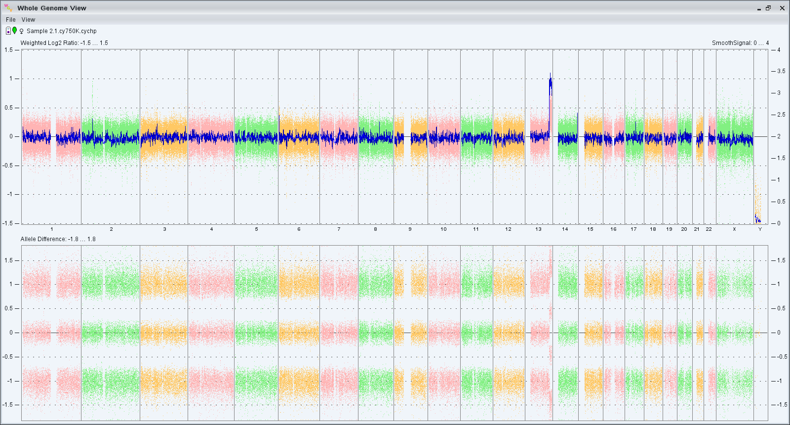
How can we study chromosomal anomalies?
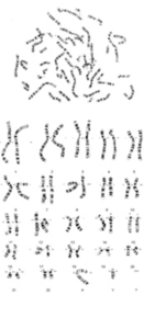
A conventional karyotype represents the arrangement of chromosomes in homologous pairs according to size, shape, and banding pattern. Karyotype analysis allows for the identification of chromosomal anomalies responsible for producing genetic diseases. Chromosome pairs 1 to 22 are called autosomes, and pair 23 consists of the sex chromosomes, called heterosomes. There are two different sex chromosomes, X and Y. Women have two X chromosomes (XX), and men have one X chromosome and one Y chromosome (XY).
Through this method we can detect all numerical anomalies of the chromosomes, but also structural anomalies larger than 5Mb. Additionally, the conventional karyotype is the only genetic diagnostic method that can detect balanced chromosomal changes: e.g., translocations.